
Magnetic resonance imaging (MRI)
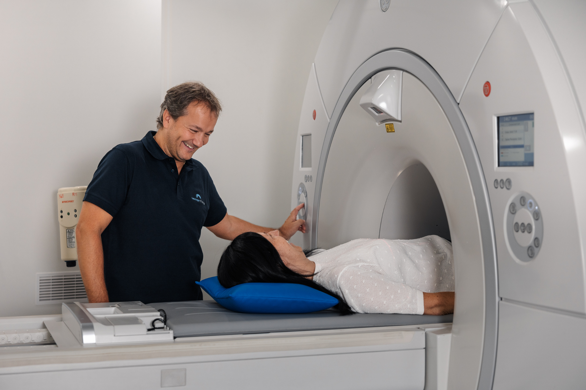
Cross-sectional images without X-ray radiation. Our 1.5-tesla scanner provides detailed images of soft tissues, joints, the spine and the brain.
Good to know
Appointment required
Book by phone:
+43 5442 64404
Current waiting time
Current waiting time for routine appointments: approximately 2–3 weeks.
Avoid missed appointment charges
If you miss an appointment without cancelling in good time, we will need to charge a missed appointment fee. If you cannot attend, please telephone us as early as possible to cancel.
Uses
When is this examination used?
MRI is particularly suitable for assessing soft tissues, joints and nerves. We use it to investigate symptoms affecting the spine and discs, as well as the knee, shoulder, hip and ankle. It also provides detailed images of the brain and skull base, and of the abdominal and pelvic organs. Breast MRI complements breast assessment, while MR angiography shows blood vessels without X-rays. MRI is particularly valuable for sports injuries, neurological conditions and planning surgery. Modern imaging sequences also help patients with claustrophobia.
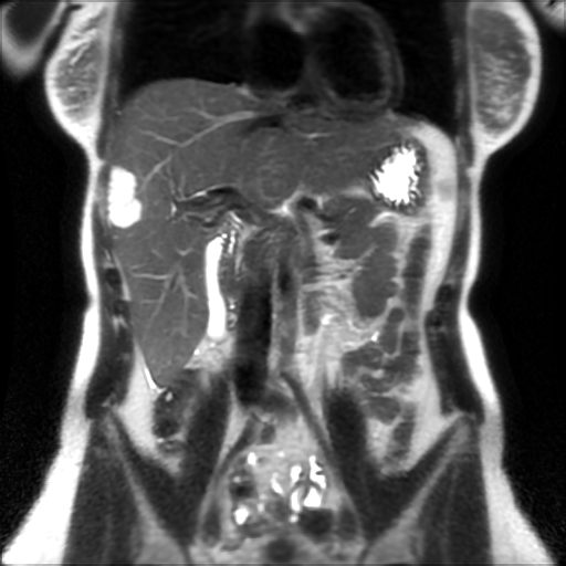
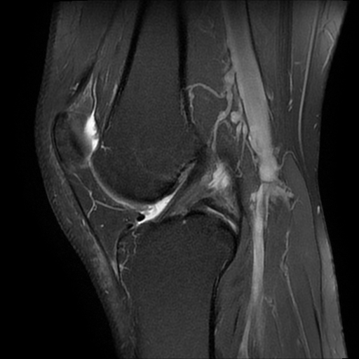
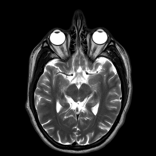
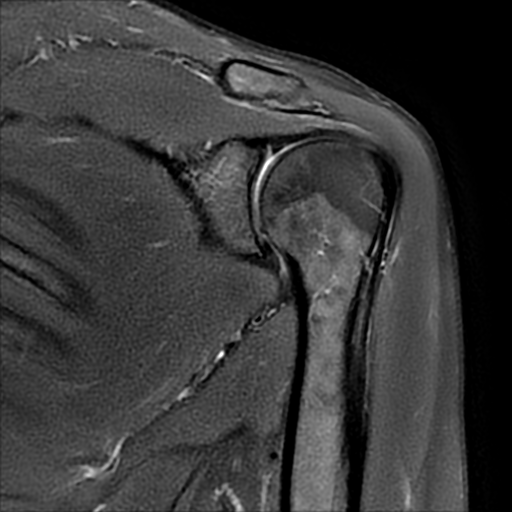
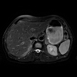
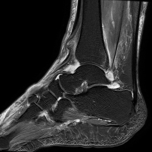
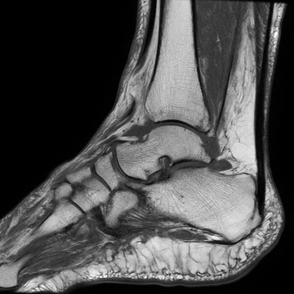
MRI image of the liver
What happens during your examination
Frequently asked questions
What should I do if I have metal in my body?
Please tell us in advance about pacemakers, implants or operations involving metal. We will check whether you can have the examination.
I have claustrophobia – what can I do?
A short general anaesthetic is possible. Please do not drive after the examination.
Will I need contrast medium?
We decide whether contrast medium is needed based on the clinical question and discuss this with you beforehand.
Does this examination involve radiation?
No. MRI uses a strong magnetic field and radio waves, with no X-rays at all.
Is this examination painless?
Yes. You lie still in the scanner. The knocking sounds can be distracting, so we provide hearing protection. Depending on the area being scanned, the examination takes 15 to 45 minutes.
How should I prepare for this examination?
Usually, no special preparation is needed. You will remove all metal objects before the examination. For abdominal MRI scans, please do not eat for a few hours beforehand.
Common examinations
- Slipped disc
- Sports injuries
- Multiple sclerosis – monitoring
- Investigation of soft tissue tumours
- Investigation of unexplained pain
Our equipment
GE 1.5-tesla MRI scanner
- High-resolution images
- Comfortable scanner bore
- Quiet imaging sequences
- Comfortable patient coils (textile blankets)
Consent forms
You can complete the forms at home or when you arrive at our practice.
Our other examinations
Computed tomography (CT scan)
High-resolution cross-sectional images. Our 64-slice CT scanner enables a substantially reduced radiation dose and short examination times.
Digital mammography
Breast screening and diagnostic assessment using our direct digital mammography system with a reduced radiation dose.
Digital X-ray
An established diagnostic imaging technique for the lungs, bones and joints — fully digital and low-dose. No appointment needed.
Ultrasound
A radiation-free examination of organs, blood vessels, the thyroid and soft tissues using a high-resolution, advanced ultrasound system.
DEXA bone density scan
Bone density scanning for the early detection and monitoring of osteoporosis — quick, painless and with a very low radiation dose.